Home
/ Ultrasound 30 Week Fetus Pictures : 30 Week 3d Ultrasound I Am So In Love With The Cheeks On My Sweet Boy The Ultrasound Tech Was 3d Ultrasound Ultrasound Pictures Gender Ultrasound Pictures - These ultrasound images show a normal early fetus of 9 weeks gestational age (1st trimester) and are taken via the transabdominal route.
Ultrasound 30 Week Fetus Pictures : 30 Week 3d Ultrasound I Am So In Love With The Cheeks On My Sweet Boy The Ultrasound Tech Was 3d Ultrasound Ultrasound Pictures Gender Ultrasound Pictures - These ultrasound images show a normal early fetus of 9 weeks gestational age (1st trimester) and are taken via the transabdominal route.
Ultrasound 30 Week Fetus Pictures : 30 Week 3d Ultrasound I Am So In Love With The Cheeks On My Sweet Boy The Ultrasound Tech Was 3d Ultrasound Ultrasound Pictures Gender Ultrasound Pictures - These ultrasound images show a normal early fetus of 9 weeks gestational age (1st trimester) and are taken via the transabdominal route.. 3 color print of your baby. Browse ultrasound pictures by week. 6 months pregnant ultrasound, symptoms, belly size. Feb 9, 2015 at 8:07 pm. These before and after images are from actual 3d/4d ultrasound sessions performed at pregnantsee!you will enjoy the same view of your little one and even see full motion video of your unborn baby.
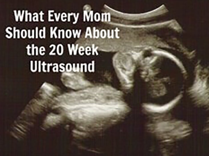
This full color medical illustration shows a fetus at the twentieth (20th) week of pregnancy. Still images from session on babyflix. Now that your unborn baby is fully formed and all of his major organ systems are functioning, most of his. 18 to 20 week ultrasound. Length, 10 1/2 inches, crown to rump;
The Anatomy Ultrasound Everything You Should Know from post.healthline.com Cutest chubby cheeks on our little one. Ultrasound of a fetus at 20 weeks. And yes the lady told me to drink 100oz a day. Moreover, baby's body proportions begin to take their natural forms. We had an ultrasound with our obgyn and filmed the experience. Your belly has grown considerably, which is probably making it very difficult for you to sleep. Collage of medical images of ultrasound anomaly scan on a female fetus 20 weeks into the pregnancy, showing child's hand, head, feet, legs, spine and human fetus age 20 weeks, illustration. The doctor regularly checks the woman's pressure, her belly circumference, determines the position of the child and the height of the uterus (with the help of ultrasound images), and constantly listens to the heartbeat of the fetus.
The baby continues to grow:
The fetus is seen, partly out of plane, under and on either side of the placenta. The purpose of this ultrasound is to be sure that your fetus is developing normally. At 30 weeks, a baby has almost completed most of her functions and all its organs are in place. Newborn baby in the egg. Week 30 comes with a myriad of symptoms, from tiredness to clumsiness.; A transvaginal ultrasound of a fetus at eight weeks. We just went yesterday at 31 weeks 4 days. An ultrasound may be performed for a variety of reasons, but looking at a baby in the womb is the most common reason. These ultrasound images show a normal early fetus of 9 weeks gestational age (1st trimester) and are taken via the transabdominal route. It is recommended that you have an ultrasound between 18 to 20 weeks. Ultrasound images at 30 weeks from this period the specialists carefully monitor all indicators of a future mother. What to expect at 19 weeks pregnant. Browse ultrasound pictures by week.
A transvaginal ultrasound of a fetus at eight weeks. This page shows typical 3d ultrasound images from 11 to 36 weeks. If you have recently had an ultrasound and you want to know how to interpret the images on your ultrasound, then you may benefit from learning about some of the basics of ultrasound imaging. Imaged using a philips hd15 system. As the baby grows, the amount of amniotic fluid will be reduced.
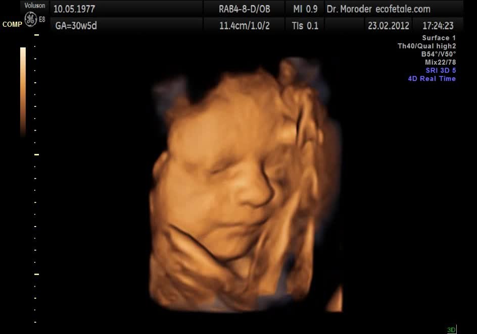
File Fetal Yawning 4d Ultrasound Ecografia 4d Dr Wolfgang Moroder Theora Ogv Wikimedia Commons from upload.wikimedia.org At this stage, the baby has put on some weight and filled out to make features more visible, yet still enough fluid in front of baby's face to obtain great images. This page shows typical 3d ultrasound images from 11 to 36 weeks. These before and after images are from actual 3d/4d ultrasound sessions performed at pregnantsee!you will enjoy the same view of your little one and even see full motion video of your unborn baby. Pregnancy symptoms at 30 weeks. An ultrasound may be performed for a variety of reasons, but looking at a baby in the womb is the most common reason. As the baby grows, the amount of amniotic fluid will be reduced. It shows the yoke sac and the umbilical cord. A scan of an ultrasound image of a 20 week old fetus.
People tell us that we have the best 3d ultrasound photo gallery comparing our 3d ultrasound photos with photos after baby's birth.we hope you will agree.
Week 30 comes with a myriad of symptoms, from tiredness to clumsiness.; At 30 weeks, a baby has almost completed most of her functions and all its organs are in place. This also shifts your center of gravity, causing you to feel unsteady on your feet — and worsening your back pain as well. And yes the lady told me to drink 100oz a day. Ultrasound scan of baby at 30 weeks gestation with description of its development at that stage 3d & 5d ultrasound images and 4d ultrasound video can be obtained at any stage. Pregnancy symptoms at 30 weeks. Ultrasound images at 30 weeks from this period the specialists carefully monitor all indicators of a future mother. Although your belly might make you feel like you have watermelon inside, the baby's height is around 15 inches (38 cm). The doctor regularly checks the woman's pressure, her belly circumference, determines the position of the child and the height of the uterus (with the help of ultrasound images), and constantly listens to the heartbeat of the fetus. The purpose of this ultrasound is to be sure that your fetus is developing normally. We had an ultrasound with our obgyn and filmed the experience. An ultrasound may be performed for a variety of reasons, but looking at a baby in the womb is the most common reason.
And yes the lady told me to drink 100oz a day. Listen to your baby's heartbeat. What to expect at 19 weeks pregnant. If you have recently had an ultrasound and you want to know how to interpret the images on your ultrasound, then you may benefit from learning about some of the basics of ultrasound imaging. The fetus is seen, partly out of plane, under and on either side of the placenta.
The Anatomy Ultrasound Everything You Should Know from post.healthline.com At 30 weeks, a baby has almost completed most of her functions and all its organs are in place. Imaged using a philips hd15 system. Still these last few weeks will add strength to a fetus and complete all its body functions so that it will be ready to arrive in this world and survive on its own. See how your baby is developing in each week of pregnancy with these ultrasound images, from weeks four to 40. Your belly has grown considerably, which is probably making it very difficult for you to sleep. The fetus is seen, partly out of plane, under and on either side of the placenta. At 30 weeks pregnant your baby is about the size of a head of cabbage. And yes the lady told me to drink 100oz a day.
Beyond the eighth week of pregnancy, the embryo is called a fetus.
Moreover, baby's body proportions begin to take their natural forms. A transvaginal ultrasound of a fetus at eight weeks. At 30 weeks, a baby has almost completed most of her functions and all its organs are in place. 3 color print of your baby. Beyond the eighth week of pregnancy, the embryo is called a fetus. The baby continues to grow: A scan of an ultrasound image of a 20 week old fetus. The placenta, the gray mass in the center of the image, as seen on a fetal ultrasound. Although your belly might make you feel like you have watermelon inside, the baby's height is around 15 inches (38 cm). See more ideas about 4d ultrasound pictures, ultrasound pictures, 4d ultrasound. Browse ultrasound pictures by week. Now that your unborn baby is fully formed and all of his major organ systems are functioning, most of his. The doctor regularly checks the woman's pressure, her belly circumference, determines the position of the child and the height of the uterus (with the help of ultrasound images), and constantly listens to the heartbeat of the fetus.